





We have already seen that hemolytic anemias result from the destruction of red blood cells (RBCs) due to external causes or internal abnormalities.
Of these, the most common example is favism, previously mentioned as a hemolytic anemia (HA) that occurs in some men after eating fava beans. This disease is caused by an inherited deficiency—linked to a gene on the X chromosome—of the enzyme glucose-6-phosphate dehydrogenase (G6PD), which is responsible for producing a substance that prevents the harmful effects of oxidative substances that, if left unneutralized, destroy red blood cells. Without this substance, whenever medications or foods with high oxidative potential—such as fava beans—are ingested, red blood cells are destroyed, leading to jaundice.
The disease manifests only in males because, unlike females, they have only one X chromosome, and the mutation present on it is not counteracted by its Y chromosome counterpart. The diagnosis is made by a clinical pathologist through measurement of the G6PD enzyme and microscopic examination of red blood cells. Hereditary abnormalities of hemoglobin (the protein that carries oxygen) are also responsible for A.H. in both sexes.
In sickle cell anemia, the presence of hemoglobin S (a slight variation of normal hemoglobin) is sufficient to deform the red blood cells, giving them a sickle-like appearance (drepnos in Greek, falce in Latin) instead of their usual disc-shaped form.
This deformation becomes more pronounced as the oxygen content in the blood decreases (low-oxygen environments, vigorous exercise, lung disease, etc.) and leads to their premature destruction because, due to their lack of flexibility, they cannot circulate through small-diameter blood vessels (capillaries), where, due to their fragility, they rupture easily, obstructing the vessels and thus causing not only acute hemolysis but also multiple small thrombi and impaired blood flow to vital organs and tissues. These factors lead to crises with various symptoms: severe abdominal or bone pain, joint pain and damage, fever, shortness of breath, chest pain, and even stroke.
The disease affects only Black people or their descendants and is diagnosed through laboratory identification of Hemoglobin S. Other forms of sickle cell anemia resulting from alterations in the alpha or beta chains of hemoglobin include alpha thalassemia (common in the Black population) and beta thalassemia (common in Mediterranean populations or those of Indian descent).
Classified as “major” or “minor” based on severity, these conditions are diagnosed in the laboratory based on the characteristics of red blood cells and the identification of the type of hemoglobin.
If you have any questions, please contact us.
We'll be happy to help.
CLINICAL PATHOLOGY LABORATORY

Related articles:
Anemia (II) - Anemia and Iron
The production of red blood cells (RBCs) in the bone marrow depends on the presence in the body of essential substances such as iron, vitamin B12, and folic acid.
Read more
Anemia Treatment: Options and How It Works
Treatment for anemia depends on the cause and may include dietary changes, supplementation, or specific medical care. The main goal is to correct the deficiency causing the anemia and restore adequat…
Read more
Anemias (I)
Driven by the heart, blood carries substances essential to life to every cell in the body.
Read more
Anemias (III) - Vitamin B12 and Anemia
In addition to iron, a deficiency in vitamin B12 (B12) is one of the causes of anemia.
Read more
Anemias (IV) - Hemolytic Anemias
I'm sure you've heard of someone who can't eat fava beans.
Read more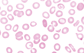
Iron Deficiency (Anemia)
Iron deficiency is a public health problem; it is the leading cause of anemia worldwide and the most prevalent nutritional deficiency.
Read more
Symptoms of Anemia: How to Recognize the Signs
Symptoms of anemia may include fatigue, weakness, paleness, and shortness of breath, which occur when the body does not receive enough oxygen through the blood. These symptoms may develop gradually a…
Read more
Types of Anemia: Learn About the Differences and Causes
The types of anemia vary depending on the cause and how they affect the production or function of red blood cells. Although anemia is often associated with iron deficiency, there are different types …
Read more